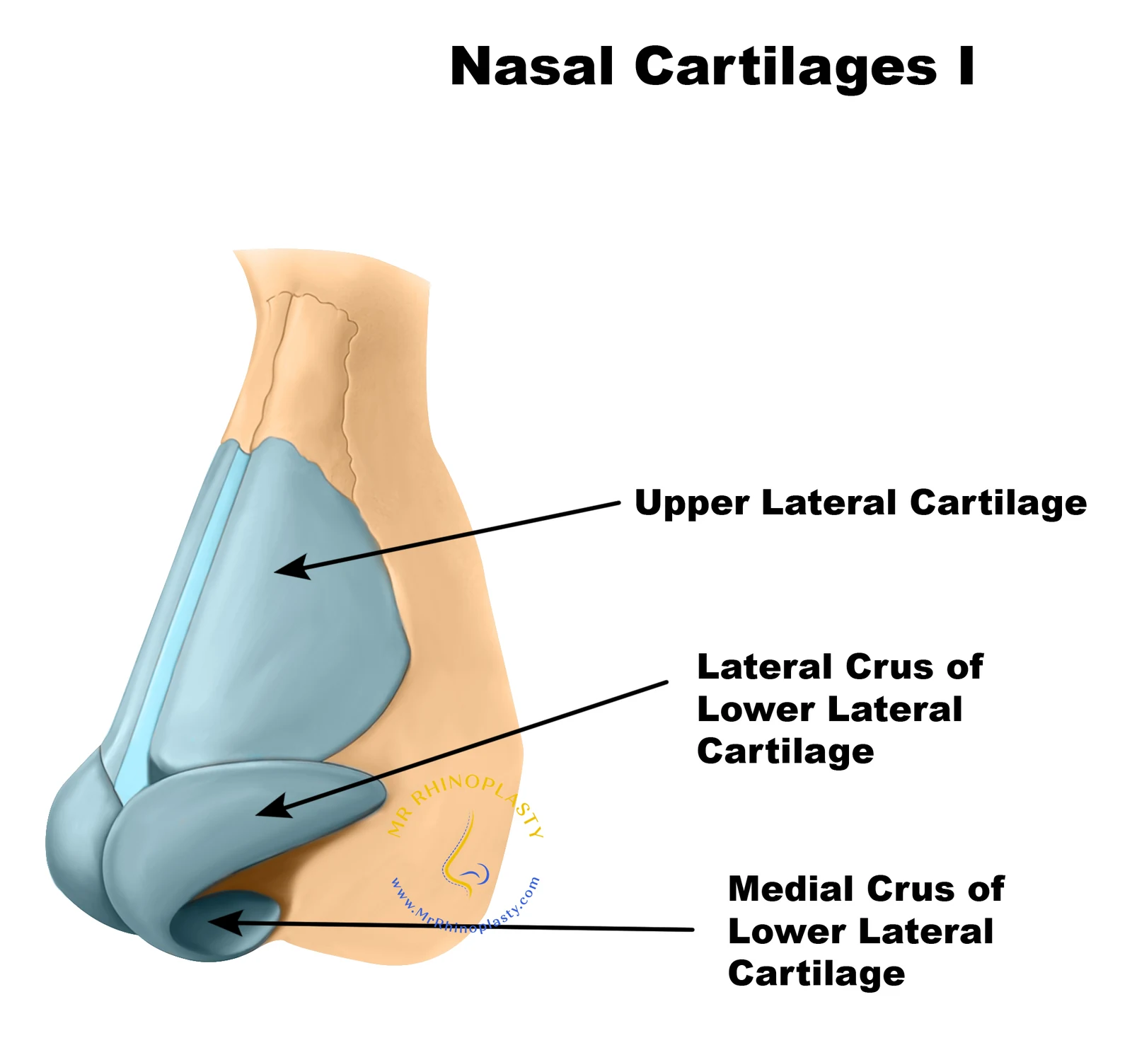
The nose is built from bone, cartilage, soft tissue and skin. The nasal bones support the upper third, the upper lateral cartilages the middle third, and the lower lateral cartilages shape the tip. Inside, the septum divides the airway and the nasal valves are its narrowest points - which is why changing the shape of the nose almost always affects how it works too.
What Are the External Parts of the Nose?
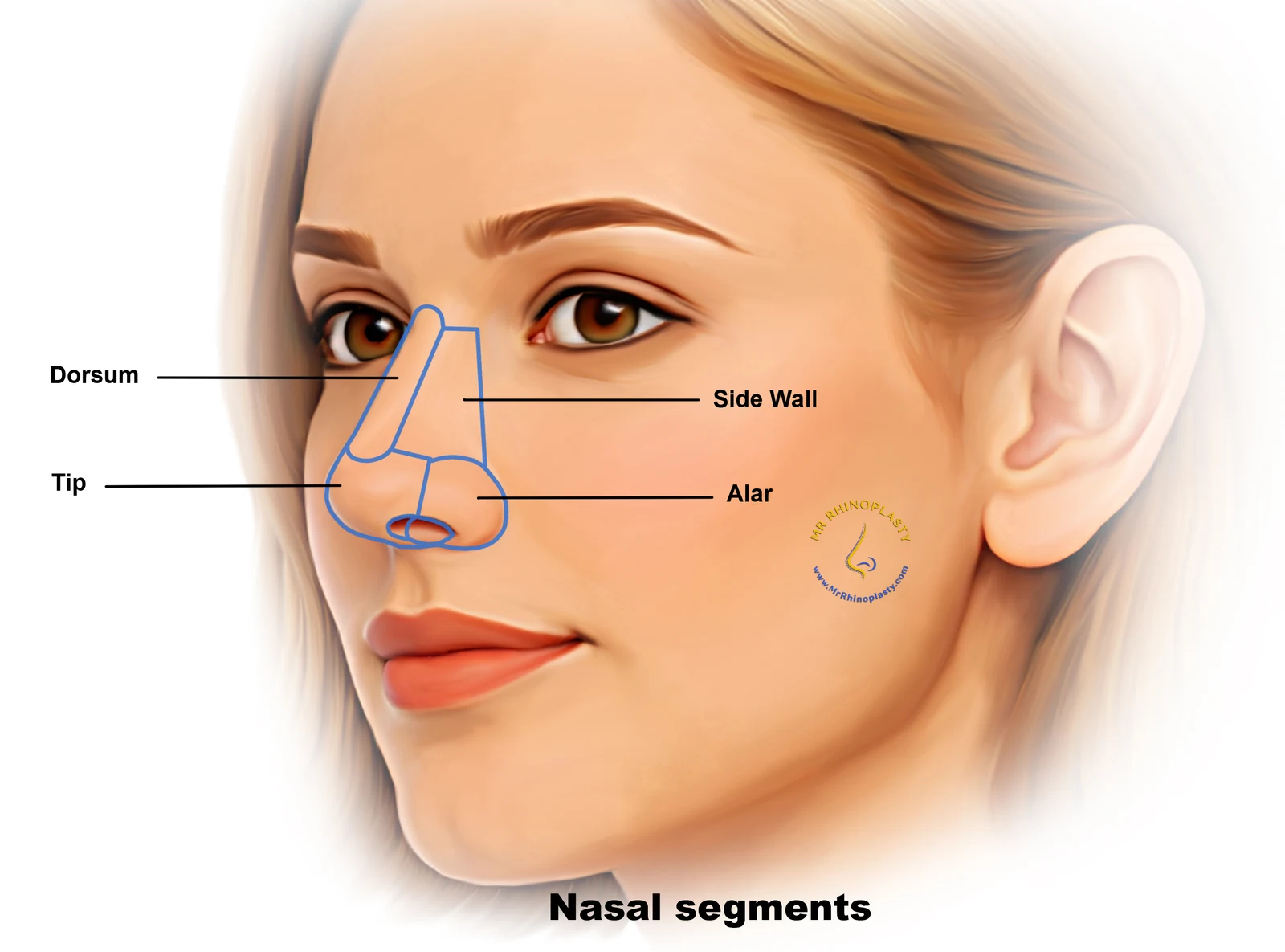
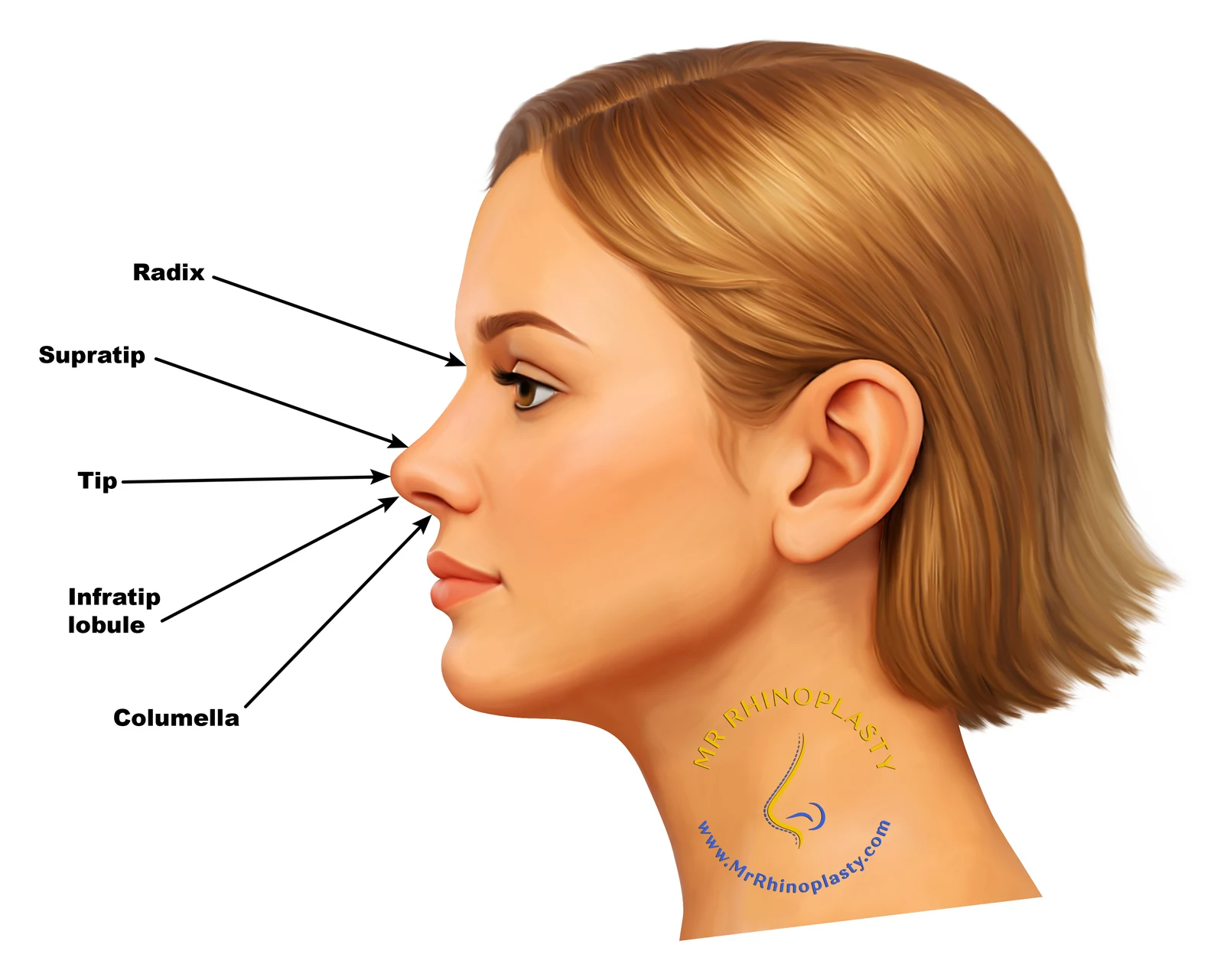
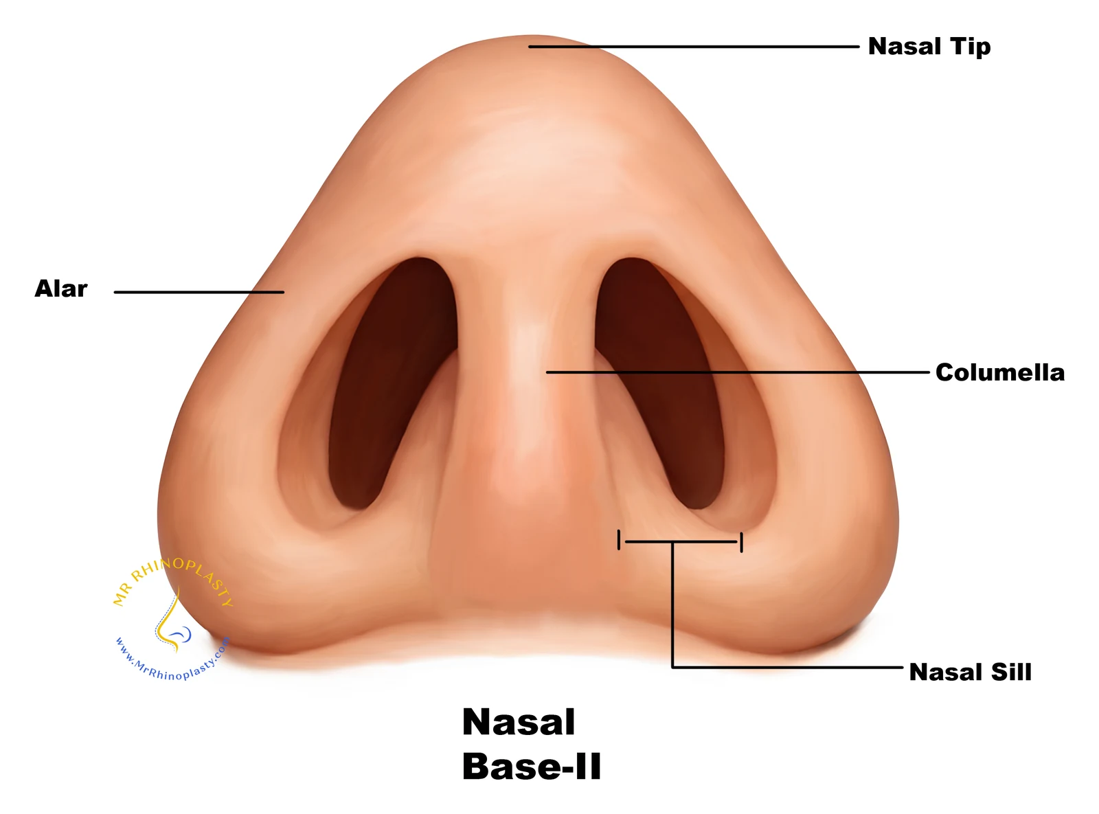
The visible parts of the nose are formed by a combination of bone and cartilage, covered by skin of varying thickness. The upper third - the bridge, or dorsum - is supported by the nasal bones, while the middle third is formed by the upper lateral cartilages. The lower third, including the tip, is shaped by the lower lateral (alar) cartilages, which determine the tip’s projection, rotation and definition. The columella is the strip of skin and cartilage separating the two nostrils, and the ala form the outer walls of the nostrils themselves. Each of these structures can be individually assessed and, where appropriate, refined during rhinoplasty.
Nasal bones
Support the upper third - the bridge, or dorsum.
Upper lateral cartilages
Form the middle third of the nose.
Lower lateral (alar) cartilages
Shape the tip: its projection, rotation and definition.
Columella
The strip of skin and cartilage separating the two nostrils.
Ala
The outer walls of the nostrils.
What’s Inside the Nose? Internal Structure and the Airway
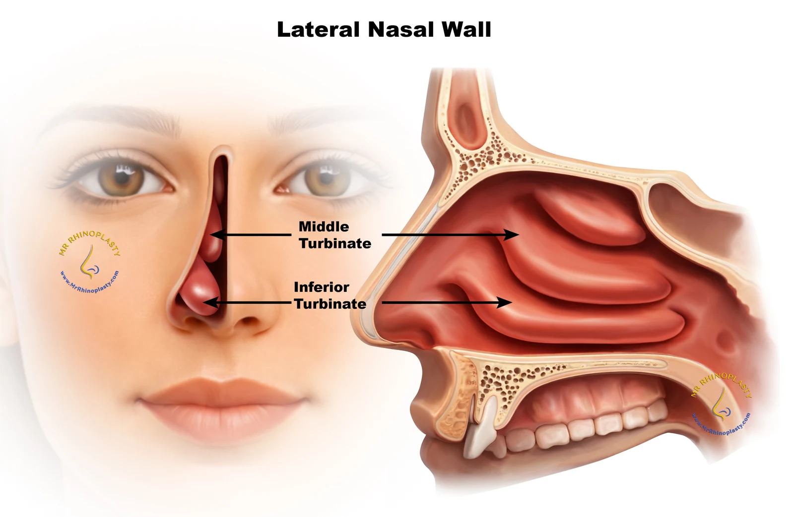
Internally, the nose is divided into two passages by the nasal septum - a wall of cartilage and bone that also provides central structural support for the entire nose. Along the side walls of each passage sit the turbinates, shelf-like structures that warm, humidify and filter the air as it passes through.
The narrowest points of the airway are the internal nasal valve (between the septum and upper lateral cartilage) and the external nasal valve (formed by the lower lateral cartilages and the rim of the nostril); collapse or narrowing at either point is a common cause of nasal obstruction, particularly during exercise or sleep. Septoplasty addresses problems with the septum itself - see What Is Septoplasty? for more detail.
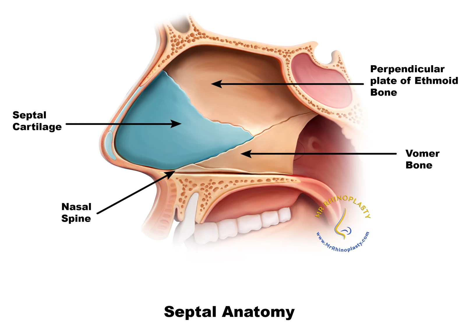
Nasal septum
Divides the nose into two passages and gives central structural support.
Turbinates
Warm, humidify and filter the air as it passes through.
Internal nasal valve
Between the septum and the upper lateral cartilage.
External nasal valve
Formed by the lower lateral cartilages and the nostril rim.
Why Does Nasal Support Matter?
The cartilage and bone framework of the nose acts as scaffolding, holding both its external shape and its internal airway open. When this support is weakened - through ageing, injury, or a previous operation - the effects are often both cosmetic and functional: the tip may drop or become pinched, the bridge may narrow, and the nasal valves may collapse, restricting airflow.
Restoring or reinforcing this support, often using the patient’s own cartilage, is central to Mr Patel’s approach to both primary and revision rhinoplasty, and reflects his particular interest in structural grafting techniques.
For more detailed information about nasal anatomy, download the free E-Book “Your Rhinoplasty Journey” from this website or watch the dedicated YouTube video about this.
Your Own Nasal Anatomy
To discuss your own nasal anatomy and how it relates to your goals, book a consultation with Mr Patel at 51 Harley Street.