Consultant surgical experience
Nasal procedures performed
Google reviews
British Society of Rhinoplasty Surgeons
Nasal valve collapse occurs when one of the narrowest points of the airway – the internal or external nasal valve – narrows further, or collapses inward, during breathing, restricting the flow of air through the nose.
The internal nasal valve is the narrowest part of the airway, between the septum and the upper lateral cartilage, while the external valve is formed by the nostril opening and its cartilage support. When the sidewall support at either point is weak, it can be drawn inwards as you breathe in, narrowing the airway further.
Nasal valve collapse is primarily a functional concern, often noticed as difficulty breathing through the nose during exercise or while lying down at night, sometimes alongside a visible pinching-in of the nostril or sidewall on breathing in.
Nasal valve collapse can be present from birth, but is also a common consequence of ageing, a previous nasal injury, or a previous rhinoplasty in which the cartilage providing sidewall support was weakened or removed.
Typical signs are a blocked nose that is worse when breathing in deeply, during exercise or at night, and a sidewall or nostril that visibly draws in on breathing. A simple test is gently pulling the cheek outwards beside the nose – if breathing improves, the nasal valve may be involved. This is assessed properly at consultation.
Nasal valve collapse is corrected by reinforcing the affected area with cartilage grafts – such as spreader grafts for the internal valve or alar batten grafts for the external valve – restoring support to keep the airway open during breathing.
Nasal valve repair is usually carried out under general anaesthetic, alone or as part of a septorhinoplasty. Spreader grafts are placed along the bridge to widen the internal valve, and batten or rim grafts strengthen the sidewalls and nostrils to support the external valve – most often using cartilage from the septum, ear or rib. The aim is a stable, open airway with a natural appearance.
Breathing usually improves progressively as internal swelling settles over the first few weeks, with further improvement over the following months. See Post Operative Care and Healing Journey and Final Results for what to expect at each stage.
If you are concerned about nasal valve collapse and its effect on your breathing, book a consultation with Mr Patel to discuss your options.
A saddle nose deformity describes a collapse or depression of the middle portion of the bridge, giving the nose a concave, or ‘scooped’, profile rather than a straight or gently curved one.
The bridge of the nose relies on the septum for support from below. When part of the septum is lost or weakened, the middle of the bridge can sink, creating a dip between the bony upper part of the nose and the tip, and in more severe cases a shortened, upturned nose.
Because the septum provides central structural support for the whole nose, a saddle nose deformity is often more than a cosmetic concern – the same loss of support that causes the bridge to collapse can also affect the airway, particularly at the nasal valves.
A saddle nose most commonly results from loss of structural support in the septum – whether through injury, previous septal surgery in which too much cartilage was removed, or, less commonly, certain inflammatory conditions affecting the cartilage. It can also follow significant trauma to the nose itself. It is also seen after cocaine or other substance abuse.
A saddle nose is most noticeable in profile as a dip or “scoop” in the middle of the bridge. Because the same loss of support can affect the nasal valves, it is often accompanied by nasal blockage, which is assessed at the same consultation alongside any underlying cause.
Correcting a saddle nose deformity involves rebuilding the lost support along the bridge, typically using cartilage grafts to restore both the height of the dorsum and the structural integrity of the septum.
Saddle nose correction is usually carried out through an open approach under general anaesthetic. Support is rebuilt with cartilage grafts – from the septum where enough remains, or more often from the ear or rib – to reconstruct the septum and restore the height and line of the bridge. Where an inflammatory condition is involved, it is important that this is stable before surgery is planned.
Most visible bruising and swelling settle over the first few weeks, while the final shape continues to refine over around 12 months as residual swelling subsides – the nasal tip is usually the last area to settle. See Post Operative Care and Healing Journey and Final Results for what to expect at each stage.

If you are concerned about a saddle nose deformity, book a consultation with Mr Patel to discuss your options.
A post-trauma nasal deformity refers to any lasting change in the shape or function of the nose following an injury, such as a fracture sustained in sport, an accident, or an assault.
An injury can shift the nasal bones, bend or fracture the septum, and damage the cartilages that support the tip and sidewalls. If these structures heal out of position, the nose can be left crooked, flattened or uneven, and the airway can be narrowed on one or both sides.
The true extent of a post-traumatic deformity is often not apparent until the swelling and bruising from the original injury have fully resolved – which can take around a week. Assessment is generally most accurate once this initial healing has settled.
Even a single significant injury can displace the nasal bones and septum, sometimes healing in a new position that affects both the external appearance of the nose and the internal airway. The nose is one of the most commonly fractured bones in the face, and not every fracture is recognised or treated at the time it occurs.
Typical features include a nose that leans to one side, a new bump or a depression along the bridge, and a blocked feeling that was not present before the injury. Many people notice both appearance and breathing changes, so both are assessed together at the same consultation.
Correcting a post-traumatic deformity often involves realigning displaced bone and cartilage, and may include septoplasty where the septum has been affected. Depending on the extent of the original injury, this can range from a relatively straightforward correction to more complex reconstruction.
Correction is usually carried out as a septorhinoplasty under general anaesthetic. The septum is straightened, displaced nasal bones are carefully realigned, and areas of collapse or irregularity are rebuilt with cartilage grafts – from the septum, ear or rib where needed. The aim is to restore both a straighter, balanced appearance and a clear airway.
Most visible bruising and swelling settle over the first few weeks, while the final shape continues to refine over around 12 months as residual swelling subsides – the nasal tip is usually the last area to settle. See Post Operative Care and Healing Journey and Final Results for what to expect at each stage.
If you are concerned about the lasting effects of a nasal injury, book a consultation with Mr Patel to discuss your options.
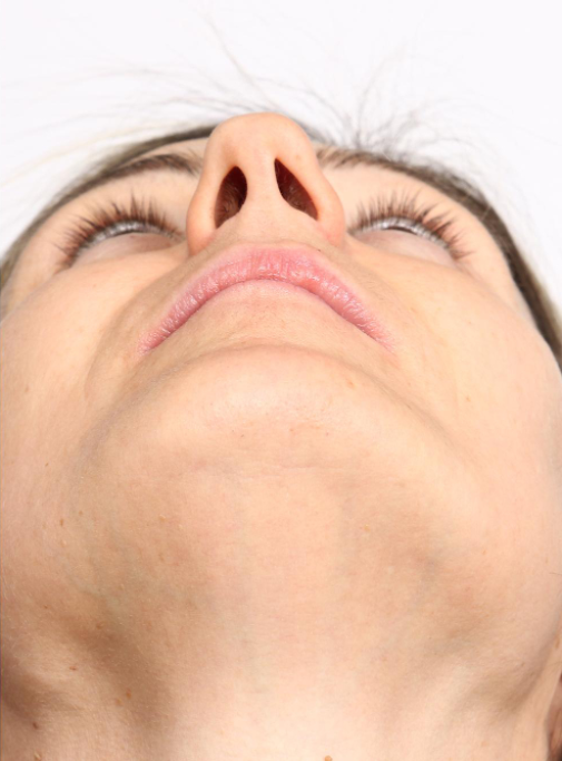
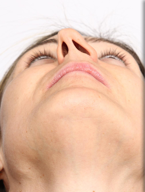
Asymmetrical nostrils describes a visible difference in the size, shape, or position of the two nostrils, most often noticed when the face is viewed from below or directly head-on.
The shape of each nostril depends on the alar cartilage on that side, the septum and columella in the middle, and the soft tissue around the nostril rim. When these differ between the left and right sides – or the septum leans to one side – one nostril can look larger, higher or a different shape from the other.
Because complete symmetry is rarely achievable – or even noticeable to others – the aim of treatment is a balanced, harmonious appearance rather than mathematically identical nostrils. This is an important part of setting realistic expectations during consultation.
Some degree of nostril asymmetry is extremely common and entirely normal – very few faces are perfectly symmetrical, and small differences between the left and right side of the nose are present in the majority of people. More noticeable asymmetry can result from a deviated septum, a previous injury, or scar tissue from earlier surgery.
Nostril asymmetry is usually most visible from below and directly from the front. Where it is caused by a deviated septum or the base of the septum dislocating into one nostril, it can be accompanied by a blocked feeling on one side, which is assessed at the same consultation.
Where nostril asymmetry is a genuine concern, correction depends on its underlying cause – ranging from septal correction to fine adjustments of the alar cartilages and surrounding soft tissue. This is assessed individually for each patient.
Correction depends on the cause and is usually carried out under general anaesthetic, often as part of a rhinoplasty or septorhinoplasty. It may involve repositioning the base of the septum, reshaping or supporting the alar cartilages, adjusting the nostril rims with small cartilage grafts, or refining the nostril bases. The aim is balanced, natural-looking nostrils rather than a perfect mirror image.
Most visible bruising and swelling settle over the first few weeks, while the final shape continues to refine over around 12 months as residual swelling subsides – the nasal tip is usually the last area to settle. See Post Operative Care and Healing Journey and Final Results for what to expect at each stage.
If you are concerned about asymmetry in your nostrils, book a consultation with Mr Patel to discuss your options.
An under-projected tip describes a nasal tip that sits close to the face, with less forward definition than is in proportion with the rest of the facial profile – sometimes appearing flat, or lacking definition, when viewed from the side.
Tip projection describes how far the tip stands out from the face. When the tip cartilages are short, small or weak, or have lost support, the tip sits closer to the face and the nose can look flat or lacking definition from the side.
An under-projected tip is different from a droopy tip: under-projection relates to how far the tip extends from the face, while droopiness relates to the angle of the tip. The two are assessed separately, although they can occur together.
Under-projection may be a natural feature of nasal anatomy, particularly where the lower lateral cartilages are smaller or weaker than average. It can also result from a previous rhinoplasty in which too much tip support or cartilage was removed.
An under-projected tip is most noticeable in profile, and can make the bridge look relatively prominent or the nose appear short and rounded. Where it follows previous surgery, it may come with other signs of weakened support, which is assessed at the same consultation.
Increasing tip projection generally involves adding structural support using cartilage grafts – often taken from the septum or rib – to bring the tip forward and create better definition.
Increasing tip projection is usually carried out as part of a rhinoplasty under general anaesthetic, most often through an open approach. Cartilage grafts – such as a columellar strut or septal extension graft, taken from the septum, ear or rib – are used to support and lengthen the tip, and tip grafts can add definition. The tip is brought forward in proportion with the bridge and the rest of the face.
Most visible bruising and swelling settle over the first few weeks, while the final shape continues to refine over around 12 months as residual swelling subsides – the nasal tip is usually the last area to settle. See Post Operative Care and Healing Journey and Final Results for what to expect at each stage.

If you are concerned about the projection of your nasal tip, book a consultation with Mr Patel to discuss your options.
A wide bridge describes a nose where the bony or cartilaginous middle section appears broad in relation to the rest of the face, most noticeable when the face is viewed head-on.
The upper part of the bridge is formed by the paired nasal bones and the middle part by the upper lateral cartilages. When these structures are set wide apart or splay outwards, the bridge looks broad from the front, and the nose can appear heavier than the rest of the face.
A wide bridge is different from a dorsal hump: width is seen when looking at the nose from the front, while a hump is seen in profile. The two are frequently assessed and treated together, but are not the same thing.
A wide bridge is often a natural feature of nasal anatomy, sometimes more pronounced in certain ethnic backgrounds or following growth during adolescence. It can also occur after a nasal fracture that widens the underlying bony framework.
A wide bridge is seen most clearly from the front and is often accompanied by a dorsal hump or a wide, heavy-looking tip. It is mainly a cosmetic concern, although where it follows a fracture, the same injury can also affect the septum and breathing, which is assessed at the same consultation.
A wide bridge can be narrowed by carefully repositioning the nasal bones – a technique often combined with dorsal hump reduction, where the two concerns frequently occur together.
Narrowing a wide bridge is usually carried out as part of a rhinoplasty under general anaesthetic. The nasal bones are carefully freed and moved inwards (osteotomies) so the bridge becomes narrower and better proportioned, and the middle cartilage vault is adjusted so the transition from bone to cartilage stays smooth. The width of the bridge is balanced with the tip and the face as a whole.
Most visible bruising and swelling settle over the first few weeks, while the final shape continues to refine over around 12 months as residual swelling subsides – the nasal tip is usually the last area to settle. See Post Operative Care and Healing Journey and Final Results for what to expect at each stage.

If you are concerned about the width of your nasal bridge, book a consultation with Mr Patel to discuss your options.
Wide nostrils refer to the width of the nasal openings at the base of the nose, sometimes described by patients, as most noticeable when the face is viewed from below or from the front. When the sides of the nose are wider than the vertical lines arising form the middle corners of the eyes, we describe this as excess alar base width. Often the nostrils widen when smiling.
The width of the base of the nose is set by the alae – the curved sides of the nostrils – and the point where they join the cheek. When the alae flare outwards or attach further apart, the base of the nose looks wide from the front and the nostril openings look large from below.
Some patients are entirely happy with the width of their nostrils and are seeking changes elsewhere in the nose; for others, narrowing the nostrils is an important part of achieving the overall balance they are looking for. Both are entirely valid starting points for a consultation.
Nostril width is largely determined by the shape and thickness of the alar cartilages and surrounding soft tissue, and varies naturally across different nasal shapes, facial proportions, and ethnic backgrounds. Wide nostrils are a normal anatomical variation, not a sign of any underlying problem.
Wide nostrils are most noticeable from the front and from below, and often more so when smiling. They are a cosmetic concern rather than a breathing problem, and the aim of any change is to bring the base of the nose into balance with the bridge, tip and the rest of the face.
Where wide nostrils are a concern, alar base reduction can narrow the width of the nostrils at their base, usually performed alongside other tip or bridge refinements.
Alar base reduction removes a small, precisely planned wedge of skin and tissue where the nostril meets the cheek, or from inside the nostril sill, to narrow the base of the nose and reduce flaring. It can be performed under local or general anaesthetic, alone or as part of a rhinoplasty. The incisions are placed in the natural crease at the base of the nostril so that they heal discreetly.
Most visible bruising and swelling settle over the first few weeks, while the final shape continues to refine over around 12 months as residual swelling subsides – the nasal tip is usually the last area to settle. See Post Operative Care and Healing Journey and Final Results for what to expect at each stage.
If you are considering changes to the width of your nostrils, book a consultation with Mr Patel to discuss your options.
A droopy tip describes a nasal tip that points downward, often becoming more noticeable when smiling, due to the angle between the tip of the nose and the upper lip – known as the nasolabial angle – being lower than average.
The position of the nasal tip depends on the strength of the tip cartilages, the ligaments that link them to the rest of the nose, and the support provided by the septum. When this support is weaker, the tip can sit lower, reducing the angle between the nose and the upper lip.
A droopy tip is different from an over-projected tip: droopiness relates to the angle of the tip, while projection relates to how far the tip extends from the face. A nose can have one of these features, both, or neither.
A droopy tip can be present from an early age as a feature of natural anatomy, but often becomes more pronounced with age, as the cartilage and supporting ligaments of the nose gradually weaken and lose some of their tension. A droopy tip can often be exacerbated when the patient smiles, due to a combination of factors, which include activity within the depressor septi muscle, which runs along the columella and can pull the tip down during smiling.
A droopy tip is most noticeable in profile and can make the nose look longer than it is. It is mainly a cosmetic concern, but because the tip and nostrils share the same support, a weak, low tip can sometimes be associated with a feeling of reduced airflow, which is assessed at the same consultation.
A droopy tip is corrected through tip rotation and support techniques, which lift the tip and improve its angle, often using a cartilage graft to maintain the new position over the long term.
Droopy tip correction is usually carried out as part of a rhinoplasty under general anaesthetic. The tip cartilages are repositioned and secured with sutures to rotate the tip upwards and improve the nose-to-lip angle, and a small cartilage graft – often from the septum – is commonly used to strengthen the tip so it keeps its new position. Where the depressor septi muscle pulls the tip down on smiling, this can be released at the same time.
Most visible bruising and swelling settle over the first few weeks, while the final shape continues to refine over around 12 months as residual swelling subsides – the nasal tip is usually the last area to settle. See Post Operative Care and Healing Journey and Final Results for what to expect at each stage.
If you are concerned about a droopy tip, book a consultation with Mr Patel to discuss your options.
An over-projected tip describes a nasal tip that extends further forward from the face than is in proportion with the rest of the facial profile – sometimes described by patients as the nose appearing too long, or too prominent, from the side.
Tip projection describes how far the tip of the nose stands out from the face. It is created by the length and strength of the tip cartilages and their support from the septum; when these are long or strong, the tip can sit further forward than suits the rest of the profile.
Projection is assessed in the context of the whole face – what looks balanced varies from one person to another, depending on factors such as forehead, lip and chin projection. This is considered carefully during consultation, rather than aiming for a single fixed proportion.
Tip projection is determined largely by the length and angle of the lower lateral cartilages and the underlying septum. An over-projected tip is most often a natural variation in nasal anatomy, though it can also become more noticeable following an injury that lengthens or displaces these structures.
An over-projected tip is most visible in profile, where the nose can look too long or prominent and can make the chin or lips appear less defined. It is mainly a cosmetic concern and is often assessed alongside the bridge and tip angle, as all three affect how the profile looks.
Reducing tip projection involves carefully adjusting the cartilage framework of the tip to bring it into better balance with the rest of the nose and face.
Reducing tip projection is usually carried out as part of a rhinoplasty under general anaesthetic. The tip cartilages are shortened or repositioned and their attachments adjusted so the tip sits back slightly, in better proportion with the bridge, lips and chin. Care is taken to keep enough support for the tip and nostrils so that the result looks natural and breathing is protected.
Most visible bruising and swelling settle over the first few weeks, while the final shape continues to refine over around 12 months as residual swelling subsides – the nasal tip is usually the last area to settle. See Post Operative Care and Healing Journey and Final Results for what to expect at each stage.
If you are concerned about the projection of your nasal tip, book a consultation with Mr Patel to discuss your options.
A pinched tip describes a nasal tip that appears narrow or tight across its middle section, sometimes with visible indentations on either side of the tip, just above the nostrils.
The lower lateral cartilages give the tip its shape and also support the sides of the nostrils. When these cartilages are weakened, over-narrowed or partly missing, the skin over the tip can sink inwards, creating a narrow, “pinched” appearance.
Because a pinched tip is most often seen after previous surgery, it is frequently encountered as part of revision rhinoplasty rather than as a primary concern – and its correction is considered alongside any other changes needed to the nose.
A pinched tip most commonly results from a previous rhinoplasty in which too much cartilage was removed from the lower lateral cartilages, leaving them too weak to support the overlying skin. It can occur, less commonly, as a natural variation in nasal anatomy without any previous surgery.
A pinched tip is most visible from the front and in three-quarter view, often as shadows or grooves either side of the tip. Because the same cartilages support the nostril rims, a pinched tip can also be associated with collapse of the nostrils on breathing in, so breathing is assessed at the same consultation.
Correcting a pinched tip generally involves rebuilding lost support using cartilage grafts, restoring a smoother, more natural contour to the tip. See Revision Rhinoplasty for more detail on this kind of structural correction.
Pinched tip correction is usually performed through an open approach under general anaesthetic. Support is rebuilt with cartilage grafts – commonly taken from the septum, the ear or, where little cartilage remains, the rib – placed to strengthen and reshape the tip and nostril rims. The aim is a smoother, more natural tip contour that is properly supported over the long term.
Most visible bruising and swelling settle over the first few weeks, while the final shape continues to refine over around 12 months as residual swelling subsides – the nasal tip is usually the last area to settle. See Post Operative Care and Healing Journey and Final Results for what to expect at each stage.
If you are concerned about a pinched tip, book a consultation with Mr Patel to discuss your options.
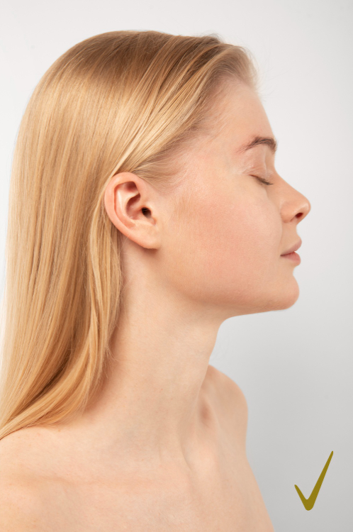
A bulbous tip describes a nasal tip that appears rounded, wide, or full, lacking the more defined contours some patients are looking for.
The shape of the nasal tip is created mainly by the two lower lateral (alar) cartilages, which curve around each side of the tip, together with the skin and soft tissue that cover them. When these cartilages are broad, rounded or set wide apart, the tip loses definition and looks full or ball-shaped.
Because skin thickness affects how much definition is achievable, the degree of refinement possible varies from patient to patient – this is discussed individually during consultation, so that expectations are realistic from the outset.
A bulbous tip is usually the result of the shape, size, or spread of the lower lateral (alar) cartilages – the paired cartilages that form the structure of the tip. The thickness of the overlying skin also plays a part; thicker skin can make the tip appear fuller, regardless of the cartilage shape beneath.
A bulbous tip is most noticeable from the front and in three-quarter view, where the tip can look wide compared with the bridge. It is a cosmetic concern rather than a breathing problem, although the tip cartilages also help support the nostrils, which is why they are reshaped carefully rather than simply removed.
Bulbous tip refinement reshapes, and where appropriate narrows, the lower lateral cartilages to create a more defined tip, while preserving enough structure to maintain a natural look and a healthy airway.
Tip refinement is usually carried out as part of a rhinoplasty under general anaesthetic, most often through an open approach for precise access to the tip cartilages. The cartilages are reshaped and narrowed with sutures and, where needed, conservative trimming, and the tip may be supported with a small cartilage graft so that it holds its new shape. In thicker skin, some soft tissue may also be carefully thinned to help definition show through.
Most visible bruising and swelling settle over the first few weeks, while the final shape continues to refine over around 12 months as residual swelling subsides – the nasal tip is usually the last area to settle. See Post Operative Care and Healing Journey and Final Results for what to expect at each stage.
If you are concerned about a bulbous tip, book a consultation with Mr Patel to discuss your options.
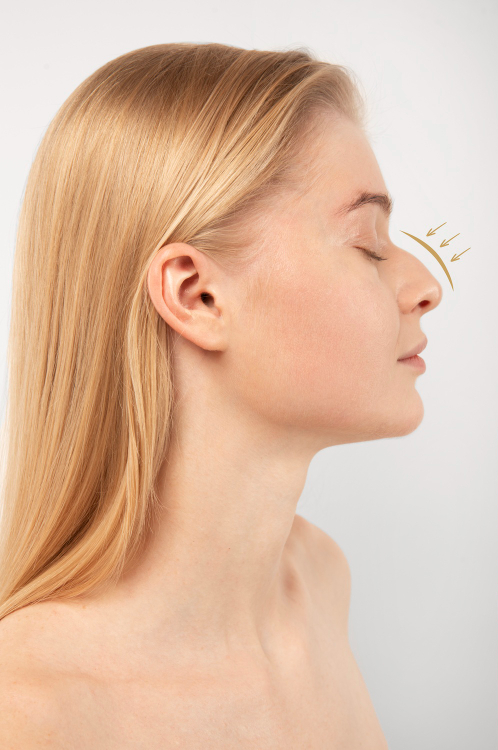
A deviated septum occurs when the wall of cartilage and bone dividing the two nostrils – the nasal septum – is displaced to one side, narrowing one or both nasal passages.
The septum is the central wall inside the nose, made of cartilage at the front and bone further back, and it supports the shape of the nose from the inside. When it bends or leans to one side, it can narrow the airway on that side – and sometimes on both sides where the bend forms an “S” shape.
Not every deviated septum requires treatment. Many people live with a deviated septum for years without symptoms, and it is often identified incidentally during examination for an unrelated concern. Treatment is generally considered when it is contributing to noticeable breathing difficulty.
Many people are born with some degree of septal deviation, while others develop it following an injury to the nose. The degree of deviation varies widely – from a minor variation that causes no symptoms, to a significant cause of nasal obstruction.
Common signs include a blocked feeling on one or both sides, difficulty breathing through the nose during exercise or sleep, snoring, and a tendency towards nosebleeds or sinus symptoms. A deviated septum may also contribute to a crooked appearance of the nose, particularly where the front part of the septum is affected.
A deviated septum is corrected through septoplasty, which straightens the septum to improve airflow. For more complex deformities, an external approach may be used. See What Is Septoplasty? for full detail, including when septoplasty is combined with rhinoplasty.
Septoplasty is usually carried out under general anaesthetic through incisions inside the nose, so there is no external scar. The bent sections of cartilage and bone are straightened, repositioned or removed, while enough support is preserved to protect the shape of the nose. Where the outside of the nose is also crooked, septoplasty can be combined with rhinoplasty in a single septorhinoplasty.
Breathing usually improves progressively as internal swelling settles over the first few weeks, with further improvement over the following months. See Post Operative Care and Healing Journey and Final Results for what to expect at each stage.
If you are concerned about a deviated septum and its effect on your breathing, book a consultation with Mr Patel to discuss your options.
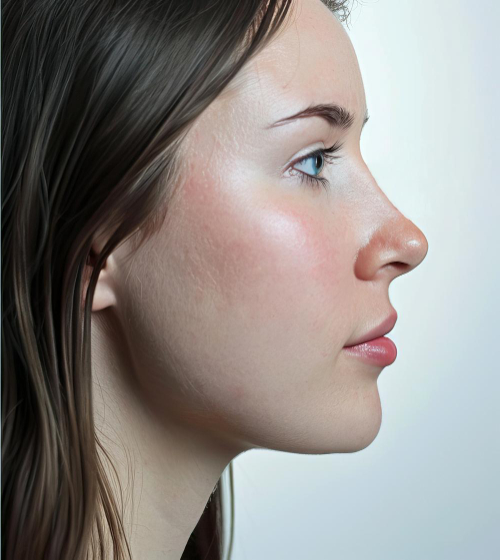
A crooked nose describes a nose that deviates to one side – along the bridge, the tip, or both – so that it no longer sits in a straight line down the centre of the face.
The nose has an upper bony framework, a middle cartilage framework and the septum running down the centre. A crooked nose occurs when one or more of these parts sit off the midline, so the bridge, the tip, or both appear to lean to one side or follow a “C” or “S” shaped curve.
Because the septum plays such a central role, a crooked nose is not always purely a cosmetic concern – deviation of the septum can also narrow the airway on one side, affecting breathing as well as appearance.
A crooked nose may be present from birth, become more noticeable as the nose grows during adolescence, or develop following an injury that displaces the underlying bone and cartilage. The nasal septum, which forms the central structural support of the nose, is very often involved in nasal deviation.
A crooked nose is most noticeable from the front, where light and shadow along the bridge can exaggerate the deviation. Because the septum is so often involved, many people with a crooked nose also notice blockage on one side, which is assessed at the same consultation.
Correcting a crooked nose involves realigning the bone and cartilage framework, often combined with septal correction (septoplasty) where the septum itself is deviated. See Crooked Nose Correction and What Is Septoplasty? for more detail.
Crooked nose correction is usually carried out as a septorhinoplasty under general anaesthetic. The septum is straightened and repositioned, the nasal bones are carefully realigned to the midline, and the middle cartilage framework may be supported with spreader grafts to keep the bridge straight. Fully straightening a nose that has been crooked for many years can be challenging, so the aim is a clearly straighter, balanced result with improved breathing.
Most visible bruising and swelling settle over the first few weeks, while the final shape continues to refine over around 12 months as residual swelling subsides – the nasal tip is usually the last area to settle. See Post Operative Care and Healing Journey and Final Results for what to expect at each stage.
If you are concerned about a crooked nose, book a consultation with Mr Patel to discuss your options.
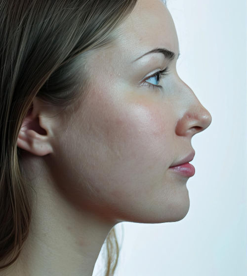
A dorsal hump is a visible bump or convexity along the bridge of the nose, most noticeable when the face is viewed from the side. It can involve the bony upper portion of the bridge, the cartilaginous lower portion, or both, and ranges from a subtle irregularity to a more pronounced curve.
The bridge of the nose – the dorsum – is supported by bone in its upper third and by cartilage below. A dorsal hump forms where this framework sits higher than the line running from the forehead to the tip, so the profile curves outwards rather than following a smooth or gently sloping line. Whether the hump is mainly bony, mainly cartilaginous or a combination of both is an important part of planning how it is corrected.
A dorsal hump is sometimes confused with a wide bridge, but the two are different: a hump refers to height or convexity seen in profile, while width is assessed when the face is viewed head-on. The two often occur together and can be addressed within the same procedure.
Dorsal humps are most often hereditary, becoming apparent once the nose finishes growing during adolescence. They can also develop, or become more noticeable, following a nasal injury – if the bone or cartilage heals with extra height along the bridge, a hump can appear where none was present before.
Because it is seen most clearly from the side, a dorsal hump can make the nose look larger or longer than it is, and can make the tip appear to point downwards. It is mainly a cosmetic concern, but it can occur alongside internal problems such as a deviated septum, so breathing is assessed at the same consultation.
A dorsal hump is corrected through dorsal hump reduction, which reshapes the bridge by removing or reducing the excess bone and cartilage to create a smoother profile in proportion with the rest of the face.
Dorsal hump reduction is usually carried out as part of a rhinoplasty under general anaesthetic. The excess cartilage is carefully trimmed and the bony part of the hump is reduced and smoothed. Where lowering the bridge leaves the top of the nose flat or wider, the nasal bones are gently repositioned so the bridge stays narrow and natural-looking. The tip and the rest of the nose are assessed at the same time, so the new profile stays balanced with the face rather than looking over-reduced.
Most visible bruising and swelling settle over the first few weeks, while the final, refined shape of the bridge continues to develop over around 12 months as residual swelling subsides. See Post Operative Care and Healing Journey and Final Results for what to expect at each stage.
If you are concerned about a dorsal hump, book a consultation with Mr Patel to discuss your options.